New bio-imaging technology peers into the heart of embryo development
Scientists using a pioneering bio-imaging system to record simultaneously the development of hundreds of aquatic embryos have discovered significant parent-offspring similarities in the timing and sequence of that development.
Researchers at Plymouth University have found the timing of key developmental milestones – such as the first beating of the heart, formation of the eyes and movement – differs markedly between individuals in a species of aquatic snail, but also that these timings appear to be heritable, i.e. they are passed from mother to offspring.
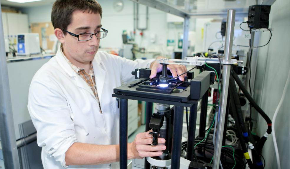
The study, made possible by a purpose-built piece of kit combining a high depth of focus lens more commonly found in aviation safety, a shutterless digital camera, and a robotic microscope stage, sheds new light on the relationship between development and evolution. It is being published in the Royal Society journal Proceedings of the Royal Society B on Wednesday 21 August.
The research, carried out in the Marine Biological and Ecology Research Centre, has taken place over the past four years as scientists carefully constructed and refined their imaging set up.
Dr Oliver Tills, project lead, said: “The link between development and evolution has remained one of the key questions in biology. Science has long suspected that heterochrony – the altered timing of developmental events between ancestors and descendants – could be the main driver of evolutionary change, but studies have primarily focused on the differences in developmental events between species with the assumption that within species variation is negligible. What we have been able to do is bring to light the astounding variation within species and show that event timing during early development is heritable.”
Snail embryos were placed in tiny little aquaria and their development recorded in high definition three-dimensional detail using time lapse photography. The bio-imaging setup enabled the team to pinpoint changes in the animal right through its body, so that they could record the precise timings of 12 key developmental milestones such as the formation of the eyes, of the shell, and hatching, spread throughout its two week embryonic development.
The team discovered marked similarities between a parent and its offspring in all 12 developmental events studied, and in two of these events – foot attachment (the transition from gliding around the egg using tiny hairs, to attaching to the inside of the egg using its muscular foot) and crawling (the same behaviour you see in an adult snail) – the strength of this similarity was sufficiently strong that it indicated heritability.
Dr Simon Rundle, Associate Professor in Freshwater Ecology, said: “There was clear evidence of similarities in the timing of all 12 physiological and morphological developmental events between parents and offspring, and of heritability in the timing of foot attachment and crawling.”
Professor John Spicer, also of Plymouth University, added: “These results challenge how we understand the interaction between development and evolution. They shed light on the potential origins of a pervasive feature in evolution – the difference in the timing of developmental events which is so evident between species. And that’s exciting.”
The research team, part of the University's Faculty of Science and Environment, and its Marine Institute, took the decision to build the unique device after they approached some of the world’s leading lens-makers and microscope manufacturers, only to be told that nothing existed that could do what they wanted. The device they’ve created has a range of between 20 to 10,000 times magnification, and can be used to study the development of species ranging from single-cell organisms to zebrafish, with up to 384 specimens in separate little aquaria at any one time.


